
Persistent Ankle Pain Is Not Always “The Old Injury”: What Should Be Reassessed?
The short answer
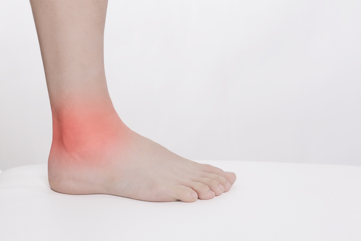
Pain that persists after an ankle sprain is not necessarily proof that the original ligament injury simply “has not healed”. Persistent ankle pain can involve bone or cartilage, tendon, impingement, ligament or stability, tarsal coalition, nerve or other sources—and more than one factor may coexist. The ACR 2025 chronic ankle pain update separates these clinical variants and emphasises matching imaging to the clinical question.[1]
Reassessment is not an invitation to self-diagnose from a list. It asks whether the location and character of pain have changed, whether function is declining, whether a new event or load change occurred, and whether the previous clinical hypothesis still explains the current response.
Why “the old injury” may be incomplete
After the first sprain, pain and swelling can change gait, weight-bearing and sport technique. Some people later develop persistent motion restriction, tendon overload, an intra-articular problem or chronic instability. In other people, the original injury has largely settled but a new training, footwear, surface or work demand creates a different irritation.
Imaging reviews describe possible chronic or overuse-related contributors such as osteochondral lesions, tendon disorders, stress injury, impingement and nerve-related conditions.[2] This does not mean that every person with long-lasting pain has one of these lesions, or that one scan identifies a single cause. It means that unchanged labels should not replace a new clinical question.
What should be asked during reassessment?
A clinician will usually clarify:
- Where and when is the pain? Lateral, anterior, posterior, medial or elsewhere? Worse with walking, hills, running, jumping, shoe pressure or rest?
- How has the pattern changed? Persistent swelling, catching, clicking, warmth, numbness, night pain or reduced weight-bearing?
- Was there a new event? Another sprain, an abrupt training increase, different shoes or surface, or a large increase in standing work?
- What function remains? Gait, single-leg stance, heel raises, stairs, running, jumping and cutting?
- Does the previous hypothesis still fit? If the location, triggers or retest no longer match, the screening pathway should change.
These answers help decide whether to modify load and rehabilitation or consider imaging and an orthopaedic, sports-medicine, rheumatology or other specialist opinion.
Imaging should answer a question, not simply increase in quantity
The ACR guideline discusses radiographs, ultrasound, MRI and CT for different chronic ankle pain variants. The choice depends on duration, the suspected structure, trauma history and previous test results.[1] Ultrasound can be useful for dynamic assessment of more superficial tendons, ligaments and muscles; MRI is suited to deeper soft tissue, cartilage and marrow; CT provides detailed information about bone and alignment. These are general modality characteristics, not a self-prescription for a patient.[2]
For example, an osteochondral lesion review notes that radiographs may be an initial tool but can miss some lesions; CT and MRI have different strengths, and MRI signal changes such as marrow oedema can affect apparent lesion size.[3] Therefore, having an MRI does not automatically provide a diagnosis, and an imaging abnormality is not automatically the pain source.
Persistent ankle pain is not the same article as chronic ankle instability
The two may coexist, but the main questions differ. Chronic ankle instability centres on repeated giving way, recurrent sprains and dynamic control. Persistent ankle pain centres on pain duration, location, changing triggers and the need to rescreen possible sources. If pain remains unchanged, function declines or new catching and swelling appear, it should not simply be managed as “instability” forever.
BAIZE Clinical Interpretation
This is an evidence-informed reasoning example, not a diagnosis, risk predictor or fixed score.
Capacity Profile
- Structural capacity: bone, cartilage, joint, ligament, tendon and previous injury status.
- Functional capacity: weight-bearing, gait, range, strength, heel raises, jumping and sport tasks.
- Biological capacity: pain, swelling, irritability, tissue recovery and overall health.
- Recovery capacity: sleep, fatigue, work, training gaps and sport scheduling.
Load Profile
- Mechanical load: walking, stairs, running, jumping, surface, footwear and volume.
- Physiological load: fatigue, concurrent injuries or systemic factors.
- Psychological load: vigilance about the old injury, avoidance, worry or repeated symptom testing.
Based on the evidence above, a BAIZE working hypothesis could be that persistent pain reflects an original problem, a coexisting problem, a capacity–load mismatch or a combination. Reassessment and retesting are needed; “old injury” should not be treated as the confirmed single cause.
Possible decision pathway
- Build capacity: once safety boundaries are clear, restore range, strength, gait and target tasks.
- Manage load: adjust walking or running dose, footwear, surface, frequency and recovery intervals.
- Reassess: repeat pain location and trend, weight-bearing and gait, range, strength, swelling or catching and target-task response. Return to clinical assessment if the pattern is not as expected.
When should a person seek prompt medical assessment?
Do not rely on online content or self-directed training after major trauma with inability to bear weight or deformity; rapidly worsening pain; a hot, red and swollen joint with fever; a cold or discoloured foot; persistent numbness; progressive weakness; persistent night pain; or unexplained systemic symptoms. These signs do not diagnose one disease, but they justify an in-person examination.
What this means for a reader
Persistent ankle pain does not automatically mean that more tests are needed, and it does not mean indefinite rest is the only option. First describe the problem again, identify the functional limitation and review load changes; then decide whether imaging or referral would answer a specific question. A “normal” scan does not invalidate symptoms, and an “abnormal” scan does not automatically mean surgery.
FAQ
How long after an ankle sprain should I seek reassessment?
There is no universal number of days. Reassessment is reasonable when symptoms are not following an expected improvement trend, weight-bearing or function remains limited, or new swelling, catching or numbness appears.
What if the scan is normal but the ankle still hurts?
Reorganise the pain location, triggers, function and load changes, then ask a qualified clinician whether a different examination or test is needed.
What do MRI, ultrasound and CT each help with?
They visualise different structures and answer different questions. The test name should not be treated as a diagnosis or treatment prescription.[1-3]
How is persistent ankle pain different from chronic ankle instability?
Persistent pain focuses on ongoing pain and rescreening its possible sources. Chronic instability focuses on repeated giving way, recurrent sprains and dynamic control; both may be present.
Can I continue sport?
Adjust according to weight-bearing, gait, target tasks and next-day response. Follow any specialist or postoperative restrictions first.
Which symptoms need prompt care?
Inability to bear weight, deformity, rapid swelling, fever with redness or heat, persistent numbness, circulation changes, progressive weakness or persistent night pain warrant prompt assessment.
References
- Expert Panel on Musculoskeletal Imaging; Schonberger A, Bartolotta RJ, Ha AS, et al. ACR Appropriateness Criteria® Chronic Ankle Pain: Update 2025. J Am Coll Radiol. 2026;23(7):1335-1347. DOI: 10.1016/j.jacr.2026.02.006. PMID: 41817475. https://pubmed.ncbi.nlm.nih.gov/41817475/
- Fritz J, et al. MR Imaging-Ultrasonography Correlation of Acute and Chronic Foot and Ankle Conditions. Magn Reson Imaging Clin N Am. 2023. DOI: 10.1016/j.mric.2023.01.009. PMID: 37019553. https://pubmed.ncbi.nlm.nih.gov/37019553/
- van Bergen CJ, Gerards R, Opdam KT, Kerkhoffs GMMJ. Diagnosing, planning and evaluating osteochondral ankle defects with imaging modalities. World J Orthop. 2015;6:944-953. DOI: 10.5312/wjo.v6.i11.944. PMID: 26716090. https://pubmed.ncbi.nlm.nih.gov/26716090/
- Michels F, Wastyn H, Pottel H, et al. The presence of persistent symptoms 12 months following a first lateral ankle sprain: a systematic review and meta-analysis. Foot Ankle Surg. 2022;28:817-826. DOI: 10.1016/j.fas.2021.12.002. PMID: 34961654. https://pubmed.ncbi.nlm.nih.gov/34961654/
- Cao S, Wang C, Ma X, Wang X, Huang J, Zhang C. Imaging diagnosis for chronic lateral ankle ligament injury: a systematic review with meta-analysis. J Orthop Surg Res. 2018;13:122. DOI: 10.1186/s13018-018-0811-4. PMID: 29788978. https://pubmed.ncbi.nlm.nih.gov/29788978/ (open full text: https://pmc.ncbi.nlm.nih.gov/articles/PMC5964890/)
- Milos R-I, Fritz L-B, Schueller-Weidekamm C. Impingement syndrome of the ankle. Radiologe. 2017;57(4):309-326. DOI: 10.1007/s00117-017-0228-9. PMID: 28324121. https://pubmed.ncbi.nlm.nih.gov/28324121/
- Barbier O, et al. Osteochondral lesions of the talar dome. Orthop Traumatol Surg Res. 2022. DOI: 10.1016/j.otsr.2022.103452. PMID: 36273506. https://pubmed.ncbi.nlm.nih.gov/36273506/
- American College of Radiology. ACR Appropriateness Criteria® Chronic Ankle Pain: official narrative. https://acsearch.acr.org/docs/69422/Narrative/
Author & Clinical Review
Written by REPULI Clinical Team
Clinically reviewed by David Konrad Lehr, PT
Master's Degree in Rehabilitation and Healthcare Management
Clinical focus: sports injury rehabilitation, post-operative functional restoration and musculoskeletal rehabilitation
Last evidence search: 2026-08-25
Last updated: 2026-09-01